Pediatric Elbow Fracture Treatment in Raleigh, NC
Child fell from the monkey bars with elbow pain and swelling? Supracondylar humerus fractures are the most common serious elbow injury in children — and displaced fractures are a time-sensitive surgical emergency.
What Is Pediatric Elbow Fracture?
Supracondylar humerus fractures — fractures just above the elbow joint — account for 60% of pediatric elbow fractures and are the most surgically important. They occur when a child falls on an outstretched hand, breaking the humerus just above the joint. The displaced fragment can stretch or injure the brachial artery and the anterior interosseous nerve.
Other important pediatric elbow fractures include lateral condyle fractures (the most commonly missed — requires surgery if >2mm displaced) and medial epicondyle avulsions (associated with elbow dislocations in older children).
ⓘ Pale, cold, or pulseless hand = vascular emergency. Go to the emergency room immediately. The brachial artery may be at risk. This is a time-sensitive limb threat requiring urgent surgical treatment.
Risk Factors
Pediatric elbow fractures occur from falls — common in active children.
Playground Falls
Monkey bars — classic supracondylar mechanism
Trampoline Falls
High-energy landing on outstretched hand
Cycling
Falls from bikes and scooters
Contact Sports
Blocking and collision falls
Gymnastics
Landing and dismount falls
Children Age 5–10
Peak age for supracondylar fractures
How Pediatric Elbow Fracture Progresses
No displacement. Cast immobilization. Excellent outcome.
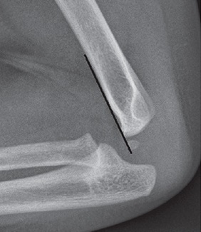
Angulated — intact posterior cortex. Closed reduction and pinning.
Complete displacement — neurovascular structures at risk. Urgent surgery.
Diagnosis
X-rays in AP and lateral views are obtained. The posterior fat pad sign on lateral X-ray indicates an occult fracture. All six ossification centers (CRITOE: Capitellum, Radial head, Internal epicondyle, Trochlea, Olecranon, External epicondyle) are assessed by expected age. Neurovascular examination is mandatory.
- ✓X-rays (AP, lateral)
- ✓Posterior fat pad sign (lateral view — occult fracture)
- ✓Neurovascular assessment — radial pulse and anterior interosseous nerve (OK sign test)
- ✓CRITOE ossification center assessment
- ✓Comparison X-ray of uninjured elbow if uncertain
Treatment Options
Dr. Chambers recommends the best approach based on your individual presentation and goals.
Long Arm Cast (Type I)
Non-displaced supracondylar fractures are treated in a long arm cast at 90° of elbow flexion for 3 weeks. Weekly X-ray at 1 week ensures no secondary displacement. Excellent outcomes reliably achieved.
Closed Reduction & Percutaneous Pinning (CRPP)
Under general anesthesia, the fracture is reduced and held with 2–3 K-wires placed through the skin. The gold standard for Type II and III fractures. Pins are removed in clinic at 3–4 weeks without anesthesia. Urgent treatment prevents neurovascular damage.
Recovery Timeline
Reduction & Pinning
Urgent surgery for Type II–III. Neurovascular confirmed after reduction. Overnight observation.
Cast Immobilization
Long arm cast. X-ray at 1 week confirms alignment. Pins protect the fracture.
Pin & Cast Removal
Pins removed in clinic without anesthesia. Elbow range-of-motion begins immediately.
Full Recovery
Full elbow motion restored. Return to sports with clearance from Dr. Chambers.
Frequently Asked Questions
It depends on the fracture type. Non-displaced (Type I) supracondylar fractures are treated with a cast — no surgery needed. Displaced fractures (Types II and III) require surgical pinning to prevent deformity and protect the nerve and blood vessel at the elbow. Lateral condyle fractures with >2mm displacement also require surgery. Dr. Chambers will evaluate the X-rays and give a clear recommendation.
The anterior interosseous nerve — a branch of the median nerve — is most commonly injured. The test is the OK sign: the child cannot make a perfect OK circle with the thumb and index finger (weak pinch). This nerve injury usually recovers fully as the fracture heals. Radial nerve injury causes weak wrist extension. Both typically recover without additional treatment.
With prompt, proper treatment, the vast majority of pediatric elbow fractures heal with excellent outcomes. The most important factor is achieving good alignment — which is why displaced fractures are treated urgently with pinning. Cubitus varus (a "gunstock" elbow deformity) from malunion is the most common complication of inadequately treated supracondylar fractures.
Related Conditions & Resources
Child With Elbow Pain After a Fall? Get Seen Today.
Displaced elbow fractures in children are time-sensitive. Same-day urgent appointments available. No referral needed.
Stephen Chambers, M.D.
Dual Board-Certified Hand & Upper Extremity Surgeon · Raleigh Orthopaedic